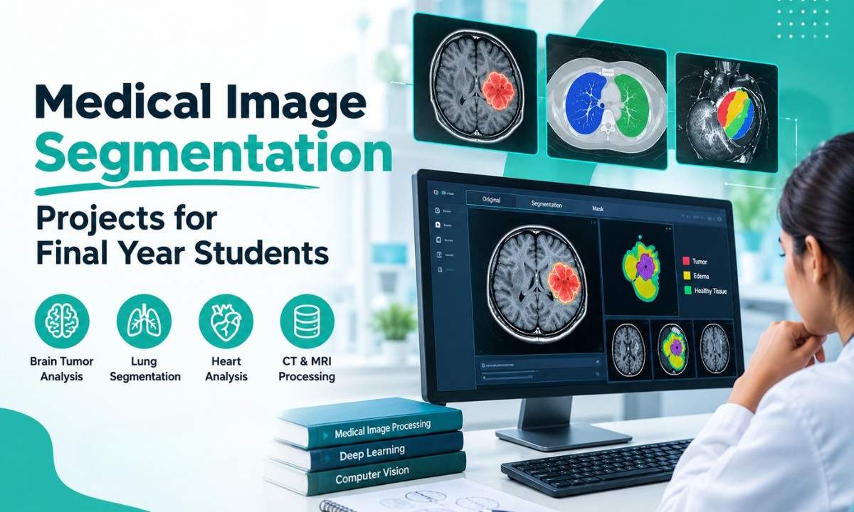

Medical Image Segmentation Projects are an excellent choice for final-year students interested in artificial intelligence, machine learning, deep learning, and medical image processing. These projects focus on identifying and separating specific regions in medical images, such as brain tumors, organs, and tissues, using MRI scans, CT images, and advanced image processing techniques.
With the growing importance of computer vision in medical research, students can explore different algorithms to analyze medical images and understand how automated segmentation works. Takeoff Edu Group helps students explore research-oriented project topics that combine image processing techniques with practical implementation. From brain tumor segmentation to wavelet-based edge detection, these Medical Image Segmentation Projects offer valuable opportunities to develop programming, analytical, and research skills.
What Is Medical Image Segmentation?
Medical image segmentation is the process of identifying and separating specific structures within medical images. For example, a segmentation algorithm can highlight a suspected tumor region in a brain MRI scan or isolate an organ from a CT image.
Unlike image classification, which assigns a label to an entire image, segmentation identifies individual pixels or voxels belonging to particular regions.
Common applications include:
- Brain tumor segmentation using MRI scans.
- Organ segmentation using CT images.
- Medical image enhancement and analysis.
- Tissue identification and boundary detection.
- Automated medical image processing research.
Students can implement these techniques using Python, OpenCV, machine learning libraries, and deep learning frameworks.

Top Medical Image Segmentation Projects for Final Year Students
The following Medical Image Segmentation Projects cover different approaches, from traditional image processing to advanced medical image analysis.
1. Brain Tumor Segmentation Using MRI Images
This project focuses on identifying and separating tumor-related regions from surrounding brain tissue in MRI scans.
Students can preprocess MRI images, apply segmentation algorithms, and visualize the identified regions using masks or overlays.
Technologies: Python, OpenCV, NumPy, and PyTorch or TensorFlow.
Learning outcome: Understand image preprocessing, segmentation algorithms, and performance evaluation.
2. MRI Brain Image Segmentation Using Thresholding
Thresholding separates image regions according to their intensity values. Students can experiment with global and adaptive thresholding methods to understand how pixel intensity influences segmentation results.
This project is suitable for beginners learning fundamental image processing techniques.
Technologies: Python, OpenCV, and NumPy.
Learning outcome: Implement thresholding algorithms and compare their effectiveness on MRI images.
3. CT Scan Image Segmentation for Organ Analysis
CT images can be used to study the segmentation of anatomical structures such as lungs, liver, and bones.
Students can develop a system that processes CT images, identifies selected structures, and visualizes the segmentation results.
Technologies: Python, SimpleITK, OpenCV, and medical imaging datasets.
Learning outcome: Understand how segmentation methods are applied to different medical imaging modalities.
4. Deep Learning-Based Medical Image Segmentation
Deep learning models can learn image features from annotated datasets and predict segmentation masks for new images.
Students can explore architectures such as U-Net for brain MRI or organ segmentation. The model can be trained using labeled images and evaluated against reference segmentation masks.
Technologies: Python, PyTorch or TensorFlow, and medical imaging datasets.
Learning outcome: Gain practical knowledge of neural networks, model training, and segmentation evaluation.
5. Wavelet-Based Edge Detection for Medical MRI Images
Edge detection identifies boundaries where image intensity changes significantly. Wavelet transforms analyze image details at different scales, making them useful for studying edges in medical images.
Students can implement wavelet filters and compare their results with conventional edge detection methods.
Technologies: Python, PyWavelets, OpenCV, and NumPy.
Learning outcome: Understand wavelet transforms, edge detection, and image signal processing.

Five Research-Based Medical Image Segmentation Project Titles
Students looking for research-oriented topics can explore the following project titles.
1. A Systematic Review of Machine Learning and Deep Learning Techniques for Brain Tumor Classification, and Grading
This project investigates machine learning and deep learning methods used to classify brain tumors and study tumor grading.
Students can compare published approaches, examine MRI-based classification techniques, and identify research challenges.
Although classification and grading are different from segmentation, segmentation can support these tasks by identifying tumor regions for further analysis.
Focus areas: Literature review, machine learning, deep learning, MRI analysis, and performance evaluation.
2. Automatic Brain Tumor Tissue Detection Based on Hierarchical Centroid Shape Descriptor in T1-Weighted MR Images
This project explores shape-based methods for detecting and analyzing candidate tumor regions in T1-weighted MRI images.
Centroid-based shape descriptors represent geometric characteristics of image regions. A hierarchical approach can examine these characteristics at multiple levels.
Students can investigate preprocessing, candidate region identification, feature extraction, and detection performance.
Focus areas: MRI processing, shape descriptors, feature engineering, and image analysis.
3. Segmentation-Driven Image Registration Application to 4D DCE-MRI Recordings of the Moving Ultrasonic Babys
The supplied title appears to contain a possible typographical error in its final phrase. Students should verify the original research title before using it in a project report.
The broader topic, segmentation-driven registration of 4D dynamic contrast-enhanced MRI images, focuses on aligning medical images acquired over time.
Students can investigate how segmentation information helps guide image registration and evaluate alignment quality across image sequences.
Focus areas: Medical image registration, dynamic MRI, segmentation masks, and image alignment.
4. Efficient Implementation of Niblack Thresholding for MRI Brain Image Segmentation
Niblack thresholding is a local thresholding technique that uses neighborhood statistics, including the local mean and standard deviation, to calculate pixel thresholds.
This project focuses on implementing the algorithm efficiently and studying its effectiveness on MRI brain images.
Students can compare different neighborhood sizes and evaluate the results against other thresholding techniques.
Focus areas: Adaptive thresholding, algorithm optimization, MRI segmentation, and image processing.
5. Design of Wavelet Filter for Edge Detection of Medical MRI Images
This project investigates the design and evaluation of wavelet filters for identifying boundaries and fine details in MRI images.
Students can study different wavelet families, apply multiscale decomposition, and compare edge detection results.
Focus areas: Wavelet transforms, signal processing, edge detection, and medical image enhancement.
Technologies Used in Medical Image Segmentation Projects:
The choice of technology depends on the project's complexity and implementation requirements.

Beginners can start with Python, OpenCV, and NumPy. Students interested in advanced segmentation can explore deep learning frameworks and specialized medical imaging tools.
How to Choose the Right Medical Image Segmentation Project:

Before selecting a final-year project, consider the following factors:
- Technical skills: Choose an algorithm that matches your programming and mathematical knowledge.
- Dataset availability: Ensure suitable medical images and reference annotations are available.
- Project complexity: Start with thresholding or edge detection if you are new to image processing.
- Research value: Identify a clear problem and compare your method with existing approaches.
- Evaluation: Use appropriate metrics, such as Dice similarity coefficient and Intersection over Union (IoU), when reference segmentation masks are available.
A well-defined project with measurable outcomes is often more valuable than a complex project without a clear implementation plan.
Conclusion
Medical Image Segmentation Projects provide final-year students with opportunities to explore artificial intelligence, computer vision, and medical image processing through practical research topics. Brain tumor segmentation, MRI and CT image analysis, Niblack thresholding, and wavelet-based edge detection offer different levels of implementation complexity for students with varying technical backgrounds.
The five research-based project titles discussed in this guide can help students explore established image processing methods and identify opportunities for further investigation.
Takeoff Edu Group encourages students to explore research-oriented Medical Image Segmentation Projects that strengthen their programming, analytical, and technical skills. By selecting a suitable topic, implementing appropriate algorithms, and evaluating results systematically, students can develop meaningful final-year projects in medical image analysis.
FAQs
1. What are the best Medical Image Segmentation Projects for final-year students?
Brain tumor segmentation using MRI, CT organ segmentation, Niblack thresholding, U-Net-based segmentation, and wavelet-based edge detection are useful topics for final-year students.
2. Which programming language is used for medical image segmentation?
Python is a popular choice because it supports image processing, machine learning, and deep learning through libraries such as OpenCV, NumPy, PyTorch, and TensorFlow.
3. Can beginners develop medical image segmentation projects?
Yes. Beginners can start with thresholding, image enhancement, and edge detection before progressing to more advanced machine learning and deep learning techniques.
4. What is the difference between image segmentation and classification?
Classification assigns a category to an image, whereas segmentation identifies the pixels or voxels belonging to specific regions within the image.
5. Which metrics are used to evaluate medical image segmentation?
Common evaluation metrics include Dice similarity coefficient, Intersection over Union, precision, and recall. The appropriate metrics depend on the project objective and available reference annotations.
 YOLO-Based Object Detection Projects for Students
 Enhancing Security in 5G Device-to-Device Communication with a Secure Gale-Shapley Algorithm
 MATLAB Projects in 5G and Beyond: Beamforming, NOMA and More
 Design and Simulation of MIMO Systems Using MATLAB
 Speech and Audio Signal Processing in MATLAB: From Basics to Projects
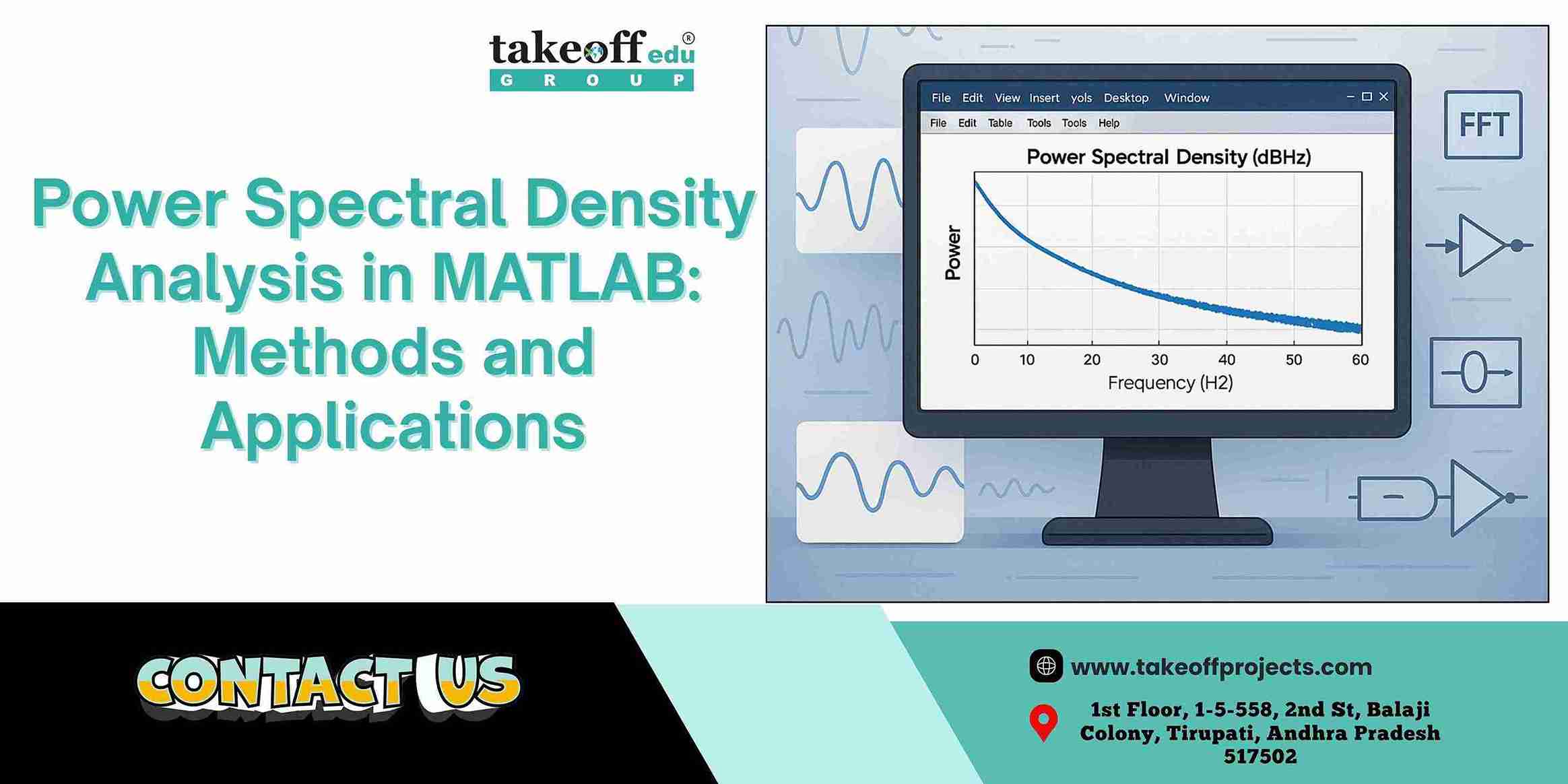 Power Spectral Density Analysis in MATLAB: Methods and Applications
 How to Filter Noise in ECG and EEG Signals Using MATLAB
 Real-Time Object Detection in Images Using MATLAB: A Practical Approach
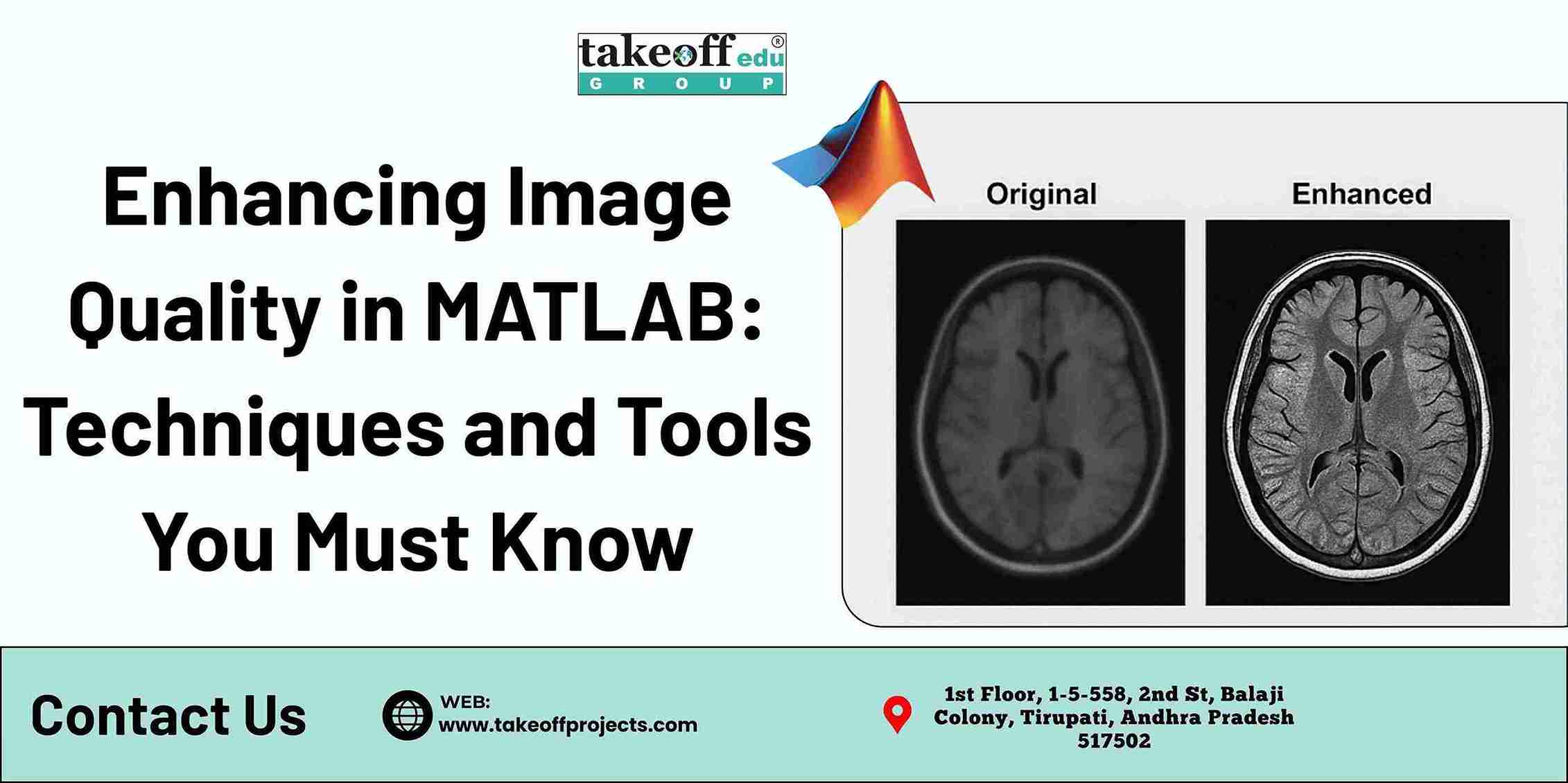 Enhancing Image Quality in MATLAB: Techniques and Tools You Must Know
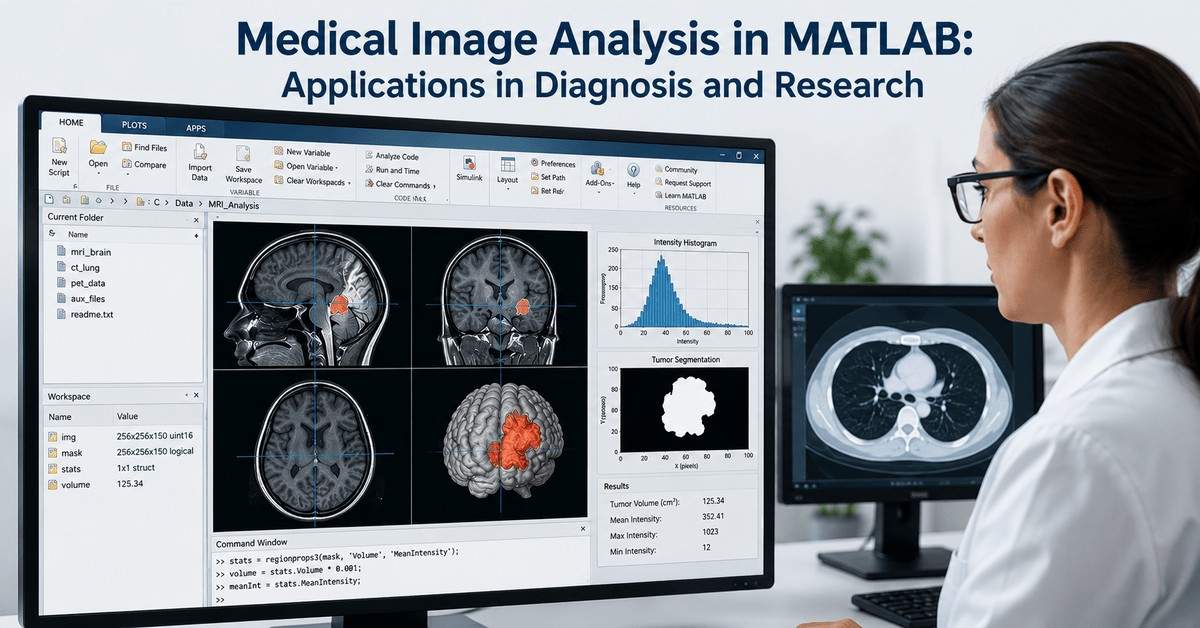 Medical Image Analysis in MATLAB: Applications in Diagnosis and Research
 Top 5 Projects on 5G Networks
 Top 5 Projects on Modulation Techniques
 Top 7 Projects on MIMO Transmissions
 Top 5 Device to Device Communications Projects
 Top 7 Image Detection Projects
 Top 7 Image Compression Projects
 Top 5 Fusion and Saliency Projects
 Top 7 Image Retrieval Projects
 Top 5 Image Denoising Projects
 Top 7 Projects on Security Applications
 Top 7 Image Enhancement Projects
 Top 10 Deep Learning Projects for B.Tech
 Top 5 Image Segmentation Projects
 Top 5 Artificial Neural Network Projects














 Paper Publishing