Automated MRI Lesion Segmentation Using Multi-Model Deep Learning Techniques
Objective
Segmentation of multiple sclerosis lesions in MRI is essential for analyzing brain abnormalities and monitoring disease progression. This project implements a multi-pathway 3D CNN for precise lesion segmentation. UNet, UNet++, and DeepLabv3 are also evaluated to compare their performance in capturing spatial and contextual features. The 3D CNN extracts volumetric information, while CRF refines boundaries for higher accuracy. A publicly available brain tumor dataset from kaggle is used to train and test the models. A Flask-based web interface allows users to upload images and visualize segmentation results, with modules for Home, Register, Login, Segmentation, and Logout. Experimental results show improved segmentation accuracy and reduced false positives.
Abstract
The segmentation of multiple sclerosis lesions in MRI images is crucial for analyzing brain abnormalities and monitoring disease progression. This project proposes a multi-pathway 3D Convolutional Neural Network (3D CNN) combined with Conditional Random Field (CRF) for automated and precise segmentation of brain lesions. The approach integrates several advanced deep learning models, including UNet, UNet++, and DeepLabv3, to extract and process spatial and contextual information efficiently. The use of 3D CNN allows capturing volumetric features of MRI images, while CRF refines the segmented output for better boundary accuracy. The project employs a publicly available brain tumor segmentation dataset to simulate lesion segmentation, enabling experimentation with different architectures and comparison of their performances. A web-based interface using Flask framework is developed for easy interaction, allowing users to upload images and visualize segmentation results. This system also includes modules such as Home, Register, Login, Segmentation, and Logout to ensure structured access and operation. Experimental results demonstrate that the proposed framework improves segmentation accuracy and reduces false-positive regions. This study contributes to automated medical image analysis by combining deep learning with probabilistic graphical models for enhanced lesion detection.
Keywords: Multiple Sclerosis, MRI, Segmentation, 3D CNN, UNet, UNet++, DeepLabv3, Conditional Random Field, Flask
NOTE: Without the concern of our team, please don't submit to the college. This Abstract varies based on student requirements.
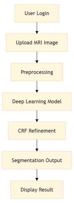
Block Diagram
Specifications
H/W CONFIGURATION:
Processor - I3/Intel Processor
Hard Disk - 160GB
Key Board - Standard Windows Keyboard
Mouse - Two or Three Button Mouse
Monitor - SVGA
RAM - 8GB
S/W CONFIGURATION:
• Operating System : Windows 7/8/10
• Server side Script : HTML, CSS, Bootstrap & JS
• Programming Language : Python
• Libraries : Flask, Pandas, MySQL. Connector, Scikit-Learn, pytorch
• IDE/Workbench : VS Code
• Technology : Python 3.8+
• Server Deployment : Xampp Server


Related Projects
 Paper Publishing

Request Call Back
Would you like to receive a free callback now?
Choose the best time for callback:
Leave your message and we will contact you as soon as possible

6-2-85/B, Old Maternity Hospital Road, Thyagaraja Nagar, Tirupati, Andhra Pradesh – 517501
+91 9030333433
+91 9393939065
0877-2261612

Disclaimer - Takeoff Edu Group Projects are not associated or affiliated with IEEE in any way. The IEEE Projects mentioned here are mentioned in the context of student projects, whose ideas are derived from IEEE publications, not projects of or by IEEE.