Segmentation of Optic Disc from Fundus Images
Abstract
This paper presents a novel method for segmentation of the Optic Disc (OD) in retinal images. Optic disc (OD) segmentation is an essential step for automated detection of various serious ophthalmic pathologies related to diabetic retinopathy. Therefore, exudates detection is our major purpose, but we must extract the OD prior to the process because it appears with similar color, intensity and contrast to other characteristics of the retinal image. The retinal image consists of blood vessels that emerge from the OD.
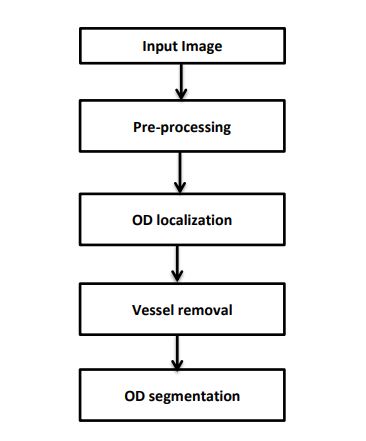
The proposed methodology includes localization of OD center, followed by elimination of vascular structure using vessel-ness filter. Finally, an active contour model was applied to boundary OD segmentation. The results are compared with a ground truth image from the ophthalmologist. The source retinal image for performing this work is obtained from the publicly available DRIVE and MESSIDOR database. This method offers a successful segmentation of OD which may help in diagnosis of various retinal abnormalities.
NOTE: Without the concern of our team, please don't submit to the college. This Abstract varies based on student requirements.
Block Diagram
Specifications

Contact Us
- info@takeoffprojects.com
- +91 9030333433, +91 9393939065


 Paper Publishing

Request Call Back
Would you like to receive a free callback now?
Choose the best time for callback:
Leave your message and we will contact you as soon as possible

6-2-85/B, Old Maternity Hospital Road, Thyagaraja Nagar, Tirupati, Andhra Pradesh – 517501
+91 9030333433
+91 9393939065
0877-2261612

Disclaimer - Takeoff Edu Group Projects are not associated or affiliated with IEEE in any way. The IEEE Projects mentioned here are mentioned in the context of student projects, whose ideas are derived from IEEE publications, not projects of or by IEEE.