Osteoporosis Detection from Dental Periapical Radiographs Using Deep Learning Approaches
Objective
The objective of this project is to develop an automated and explainable deep learning system for detecting osteoporosis from dental periapical radiographs. The system classifies images into Normal, Osteopenia, and Osteoporosis categories using Vision Transformer and Siamese Network models. It aims to provide a cost-effective and accessible alternative to DXA scans by utilizing routine dental X-rays. The project also integrates Grad-CAM++ for visual interpretability and implements a Flask-based web application for user-friendly prediction, analysis, and management of osteoporosis screening results.
Abstract
Osteoporosis is a progressive skeletal condition marked by loss of bone strength, which can lead to increased fragility and fractures. Although traditional diagnostic methods like dual-energy X-ray absorptiometry (DXA) provide accurate assessments, these methods are costly and not easily accessible for frequent screening. To address the need for affordable early detection, this project explores the use of dental radiographs as an alternative imaging source for identifying signs of systemic bone density changes. The dataset comprises annotated dental X-ray images, commonly obtained during routine dental examinations. Two deep learning approaches were implemented and evaluated: A Vision Transformer architecture with self-attention (ViT-Attn) and a Siamese network trained using a few-shot learning strategy. The ViT-Attn model captures complex spatial relationships across image regions, which is beneficial for detecting subtle patterns associated with changes in bone and dental structures. The Siamese network leverages pairwise similarity learning to perform classification effectively despite limited training samples. Both models were trained with standard pre-processing and augmentation techniques and evaluated using metrics such as accuracy, F1-score, area under the curve (AUC), and confusion matrices. Explainability was incorporated through Grad-CAM++ visualizations, providing heatmaps that highlight regions of the dental X-ray images influencing model decisions. The results demonstrate that the ViT-Attn model achieves superior performance in distinguishing between Normal, Osteopenia, and Osteoporosis cases, while the Siamese network remains robust in low-data scenarios. The study concludes that dental radiographs, when analysed with appropriate deep learning methods, can serve as an accessible screening technique for osteoporosis.
Keywords: dental radiographs, osteoporosis screening, Vision Transformer, Siamese network, deep learning, classification, explainable AI, Grad-CAM++, bone density, medical imaging.
NOTE: Without the concern of our team, please don't submit to the college. This Abstract varies based on student requirements.
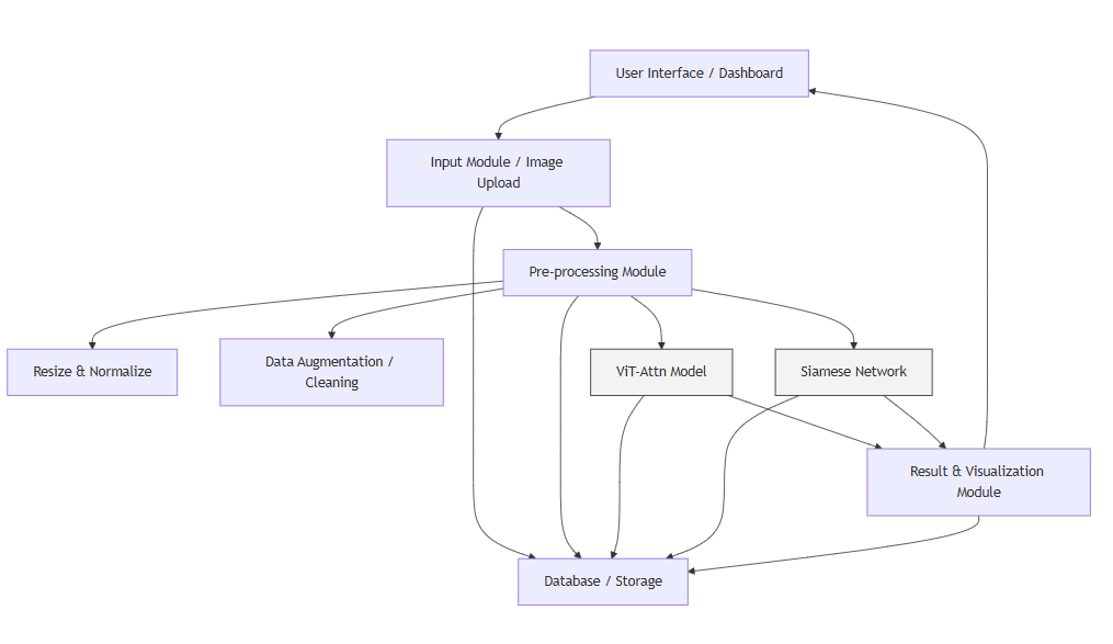
Block Diagram
Specifications
4.3 Hardware Requirements
Processor - I3/Intel Processor
Hard Disk - 160GB
Key Board - Standard Windows Keyboard
Mouse - Two or Three Button Mouse
Monitor - SVGA
RAM - 8GB
4.4 Software Requirements
Operating System : Windows 7/8/10
Programming Language : Python
Libraries : Pandas, Numpy, scikit-learn.
IDE/Workbench : Visual Studio Code.
Framework : Flask


 Paper Publishing

Request Call Back
Would you like to receive a free callback now?
Choose the best time for callback:
Leave your message and we will contact you as soon as possible

6-2-85/B, Old Maternity Hospital Road, Thyagaraja Nagar, Tirupati, Andhra Pradesh – 517501
+91 9030333433
+91 9393939065
0877-2261612

Disclaimer - Takeoff Edu Group Projects are not associated or affiliated with IEEE in any way. The IEEE Projects mentioned here are mentioned in the context of student projects, whose ideas are derived from IEEE publications, not projects of or by IEEE.