Lung Cancer Image Segmentation using Various Image Processing Techniques
Objective
The main objective of this project segmentation of Lungs CT images using different segmentation techniques
Abstract
The computer-based process of identifying the boundaries of lung from surrounding thoracic tissue on computed tomographic (CT) images, which is called segmentation, is a vital first step in radiologic pulmonary image analysis. Many algorithms and software platforms provide image segmentation routines for quantification of lung abnormalities; however, nearly all of the current image segmentation approaches apply well only if the lungs exhibit minimal or no pathologic conditions. When moderate to high amounts of disease or abnormalities with a challenging shape or appearance exist in the lungs, computer-aided detection systems may be highly likely to fail to depict those abnormal regions because of inaccurate segmentation methods. In particular, abnormalities such as pleural effusions, consolidations, and masses often cause inaccurate lung segmentation, which greatly limits the use of image processing methods in clinical and research contexts. In this review, a critical summary of the current methods for lung segmentation on CT images is provided, with special emphasis on the accuracy and performance of the methods in cases with abnormalities and cases with exemplary pathologic findings. The currently available segmentation methods can be divided into methods. The feasibility of each class and its shortcomings are explained and illustrated with the most common lung abnormalities observed on CT images. In an overview, practical applications and evolving technologies combining the presented approaches for the practicing radiologist are detailed. A CT lung image was proposed using the combination of Neural Networks and region growing which combines the regions of different pixels. The proposed approach expresses a method for segmenting the lung region from lung Computer Tomography (CT) images. This method is proposed to obtain an optimal segmented region. The first step begins by the process of finding the area which represents the lung region. In order to achieve this, the region of the entire pixel present in the entire image is grown. Second step is the grown region values are given as input to the Echo state neural networks in order to obtain the segmented lung region.
Keywords: Lungs Images, Region growing image segmentation, K means clustering image segmentation, Marker controlled watershed image segmentation.
NOTE: Without the concern of our team, please don't submit to the college. This Abstract varies based on student requirements.
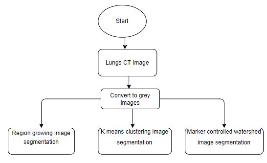
Block Diagram
Specifications
SYSTEM SPECIFICATIONS:
H/W Specifications:
- Processor : I5/Intel Processor
- RAM: 8GB (min)
- Hard Disk: 128 GB
S/W Specification:
- Operating System: Windows 10
- Server-side Script: Python 3.6
- IDE: Jupyter Notebook, VScode
- Libraries Used: Numpy, OS, pandas,Opencv
Learning Outcomes
LEARNING OUTCOMES:
- Practical exposure to
- Hardware and software tools
- Solution providing for real time problems
- Working with team/individual
- Work on creative ideas
- Testing techniques
- Error correction mechanisms
- What type of technology versions is used?
- Working of Tensor Flow
- Implementation of Deep Learning techniques
- Working of CNN algorithm
- Working of Transfer Learning methods
- Building of model creations
- Scope of project
- Applications of the project
- About Python language
- About Deep Learning Frameworks
- Use of Data Science


Related Projects
 Paper Publishing

Request Call Back
Would you like to receive a free callback now?
Choose the best time for callback:
Leave your message and we will contact you as soon as possible

6-2-85/B, Old Maternity Hospital Road, Thyagaraja Nagar, Tirupati, Andhra Pradesh – 517501
+91 9030333433
+91 9393939065
0877-2261612

Disclaimer - Takeoff Edu Group Projects are not associated or affiliated with IEEE in any way. The IEEE Projects mentioned here are mentioned in the context of student projects, whose ideas are derived from IEEE publications, not projects of or by IEEE.