Kidney Tumor Detection in CT scans Combining 3D CNN and Transformer Networks
Objective
The primary objective of this project is to develop a robust and efficient hybrid deep learning model for accurate detection and classification of kidney tumors in CT scan volumes. By integrating 3D Convolutional Neural Networks (CNNs) with transformer-based attention mechanisms, the system aims to capture both fine-grained spatial features and long-range inter-slice dependencies across multi-slice CT inputs. The goal is to enhance diagnostic accuracy, particularly in identifying malignant lesions, while ensuring computational efficiency for real-time clinical deployment. The project also seeks to provide interpretable insights through attention visualization, supporting radiologists in making informed diagnostic decisions.
Abstract
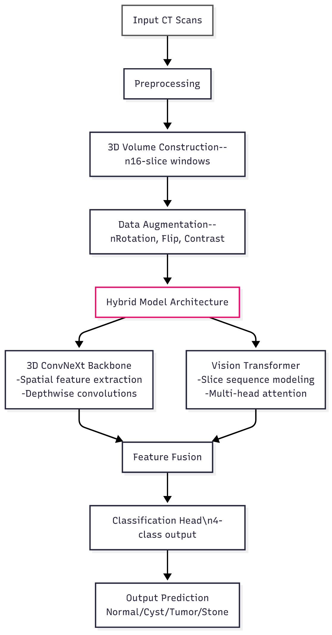
This paper introduces a hybrid deep learning architecture for kidney tumor detection in CT scans, integrating 3D convolutional operations with transformer-based sequence modeling. Our volumetric processing framework combines the local feature extraction capabilities of 3D CNNs with the global contextual understanding of transformers, specifically designed for multi-slice CT analysis. The system processes 16-slice CT volumes through a ConvNeXt-inspired 3D backbone that maintains spatial relationships across imaging planes, followed by a transformer encoder that captures long-range dependencies between slices through learned attention patterns.
The architecture employs several key innovations: (1) a depth-aware patch embedding layer that preserves inter-slice relationships when transitioning from 3D convolutions to 2D attention maps, (2) adaptive positional encodings that account for variable slice spacing in medical CT acquisitions, and (3) a multi-scale feature fusion module that combines hierarchical representations from both pathways. We demonstrate the system's clinical utility through interpretable attention visualizations that highlight diagnostically relevant regions across slice sequences.
Trained on a diverse dataset of kidney CT volumes encompassing normal anatomy and pathological findings (cysts, tumors, and stones), our approach shows particular sensitivity in detecting malignant lesions while maintaining specificity across all classes. The model's computational efficiency enables practical deployment, with inference times suitable for clinical workflows. Comparative evaluations against conventional 3D CNNs and pure transformer architectures reveal significant improvements in handling slice-to-slice variations while preserving spatial detail. This work advances volumetric medical image analysis by demonstrating how hybrid architectures can leverage both local texture patterns and global contextual information in multi-slice diagnostic imaging.
Keywords: kidney tumor detection, 3D CNN, vision transformer, CT scan classification, volumetric medical imaging, deep learning in radiology, hybrid neural networks, multi-slice analysis, ConvNeXt-Transformer fusion, diagnostic AI.
NOTE: Without the concern of our team, please don't submit to the college. This Abstract varies based on student requirements.
Block Diagram
Specifications
SOFTWARE REQUIREMENS
Operating System : Windows 7/8/10
Server side Script : HTML, CSS, Bootstrap & JS
Programming Language : Python
Libraries : Flask, Pandas, Torch, Keras, Sklearn, Numpy , Seaborn
IDE/Workbench : VSCode
Server Deployment : Xampp Server
Database : MySQL
HARDWARE REQUIREMENTS
Processor - I3/Intel Processor
RAM - 8GB (min)
Hard Disk - 128 GB
Key Board - Standard Windows Keyboard
Mouse - Two or Three Button Mouse
Monitor - Any

Demo Video


Request for Video
 Paper Publishing
Request Call Back
Would you like to receive a free callback now?
Choose the best time for callback:
Leave your message and we will contact you as soon as possible

6-2-85/B, Old Maternity Hospital Road, Thyagaraja Nagar, Tirupati, Andhra Pradesh – 517501
+91 9030333433
+91 9393939065
0877-2261612

Disclaimer - Takeoff Edu Group Projects are not associated or affiliated with IEEE in any way. The IEEE Projects mentioned here are mentioned in the context of student projects, whose ideas are derived from IEEE publications, not projects of or by IEEE.