X-ray Enhancement based on Component Attenuation, Contrast Adjustment and Image Fusion
Abstract
Inspecting X-ray images is an essential aspect of medical diagnosis. However, due to an X-ray’s low contrast and low dynamic range, important aspects such as organs, bones, and nodules become difficult to identify. Hence, contrast adjustment is critical, especially in view of its ability to enhance the details in both bright and dark regions.
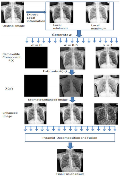
we proposed enhancing the visual contrast by adaptive tissue attenuation and dynamic range stretching. Using component decomposition and tissue attenuation, a parametric adjustment model was deduced to generate many enhanced images at once. Finally, an ensemble framework was proposed for fusing these enhanced images and producing a high contrast output in both bright and dark regions.
We have used measurement metrics to evaluate our system and achieved promising scores in each. Moreover, we applied our system to an X-ray data set provided by the Japanese Society of Radiological Technology to help with nodule detection. The experimental results of which demonstrated the effectiveness of our method.
NOTE: Without the concern of our team, please don't submit to the college. This Abstract varies based on student requirements.
Block Diagram
Specifications

Contact Us
- info@takeoffprojects.com
- +91 9030333433, +91 9393939065


 Paper Publishing

Request Call Back
Would you like to receive a free callback now?
Choose the best time for callback:
Leave your message and we will contact you as soon as possible

6-2-85/B, Old Maternity Hospital Road, Thyagaraja Nagar, Tirupati, Andhra Pradesh – 517501
+91 9030333433
+91 9393939065
0877-2261612

Disclaimer - Takeoff Edu Group Projects are not associated or affiliated with IEEE in any way. The IEEE Projects mentioned here are mentioned in the context of student projects, whose ideas are derived from IEEE publications, not projects of or by IEEE.