Performance Comparison and Analysis of Medical Image Segmentation Techniques
Abstract
This research aims at performance analysis and comparison of medical image segmentation techniques for surgical application. Existing algorithm when applied on digital images it can avoid problems such as noise and signal restoration during processing. In this we have studied CT images for performance analysis of the algorithms.This paper shows implementation and analysis of conventional Segmentation Protocol for CT Head and Neck Images. Later part of the research deals with designing an efficient protocol which does segmentation with pseudo coloring of segmented area which makes images visually efficient to extract the structure.
This designing requires three steps the pre-segmentation followed by indexing of gray level with pseudo-color map and comprehensive edge detection. Due to effective use of pseudo coloring the inside anatomical structure of craniofacial portion the noise has no effect on the segmentation results. ITK-SNAP Medical Image Segmentation Tool is used for Simulation. MATLAB is used for implementation of proposed segmentation model. The resultant images are more clear, closed, continuous, more accurate for abnormality detection.
NOTE: Without the concern of our team, please don't submit to the college. This Abstract varies based on student requirements.
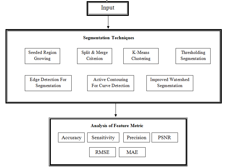
Block Diagram

Specifications

Contact Us
- info@takeoffprojects.com
- +91 9030333433, +91 9393939065


 Paper Publishing
