Novel Algorithm for Medical Image Segmentation
Objective
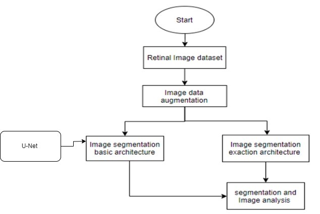
In proposed method we are performing the Retinal vessels image segmentation using the modified architecture of UNet++.
Abstract
Segmentation of retinal vessels plays a crucial role in detecting many eye diseases, and its reliable computerized implementation is becoming essential for automated retinal disease screening systems. A large number of retinal vessel segmentation algorithms are available, but these methods improve accuracy levels. Their sensitivity remains low due to the lack of proper segmentation of low contrast vessels, and this low contrast requires more attention in this segmentation process. In this paper, we have proposed new pre-processing steps for the precise extraction of retinal blood vessels. These proposed pre-processing steps are also tested on other existing algorithms to observe their impact. There are two steps to our suggested module for segmenting retinal blood vessels. The first step involves implementing and validating the pre-processing module. The second step applies these pre-processing stages to our proposed binarization steps to extract retinal blood vessels. The proposed pre-processing phase uses the traditional image-processing method to provide a much-improved segmented vessel image. Our binarization steps contained the image coherence technique for the retinal blood vessels. Retinal vessels identification and localization aim to separate the different retinal vasculature structure tissues, either wide or narrow ones, from the fundus image background and other retinal anatomical structures such as optic disc, macula, and abnormal lesions. Retinal vessels identification studies are attracting more and more attention in recent years due to non-invasive fundus imaging and the crucial information contained in vasculature structure which is helpful for the detection and diagnosis of a variety of retinal pathologies included but not limited to: Diabetic Retinopathy (DR), glaucoma, hypertension, and Age-related Macular Degeneration (AMD). With the development of almost two decades, the innovative approaches applying computer-aided techniques for segmenting retinal vessels are becoming more and more crucial and coming closer to routine clinical applications. Moreover, the evaluation and validation of the results of retinal vessels segmentation are discussed. Finally, an objective assessment is presented and future developments and trends are addressed for retinal vessels identification techniques.
Keywords: - Retinal vessels Segmentation, Retinal image dataset, Image segmentation, and segmentation algorithms.
NOTE: Without the concern of our team, please don't submit to the college. This Abstract varies based on student requirements.
Block Diagram
Specifications
H/W Specifications:
- Processor: I5/Intel Processor
- RAM : 8GB (min)
- Hard Disk : 128 GB
S/W Specifications:
• Operating System : Windows 10
• Server-side Script : Python 3.6
• IDE : PyCharm,Jupyter notebook
• Libraries Used : Numpy, IO, OS, Flask, Keras, pandas, tensorflow, Segmentation
Learning Outcomes
· Practical exposure to
· Hardware and software tools
· Solution providing for real time problems
· Working with team/individual
· Work on creative ideas
· Testing techniques
· Error correction mechanisms
· What type of technology versions is used?
· Working of Tensor Flow
· Implementation of Deep Learning techniques
· Working of CNN algorithm
· Working of Transfer Learning methods
· Building of model creations
· Scope of project
· Applications of the project
· About Python language
· About Deep Learning Frameworks
Use of Data Science


 Paper Publishing

Request Call Back
Would you like to receive a free callback now?
Choose the best time for callback:
Leave your message and we will contact you as soon as possible

6-2-85/B, Old Maternity Hospital Road, Thyagaraja Nagar, Tirupati, Andhra Pradesh – 517501
+91 9030333433
+91 9393939065
0877-2261612

Disclaimer - Takeoff Edu Group Projects are not associated or affiliated with IEEE in any way. The IEEE Projects mentioned here are mentioned in the context of student projects, whose ideas are derived from IEEE publications, not projects of or by IEEE.