MRI Breast Tumor Segmentation Using Different Encoder and Decoder Using CNN Architecture
Objective
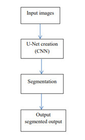
The main objective of this method is to segment the tumor part of the medical image. This segmentation is mainly depends on the CNN techniques by using Unet Architecture. By using this architecture, the tumor in the medical images can be easily segmented
Abstract
Detecting an anomaly such as a malignant tumor or a nodule from medical images including mammogram, CT, or PET images is still an ongoing research problem drawing a lot of attention with applications in medical diagnosis. The learned model can be used to classify a testing sample into a positive or negative class.
However,
in medical applications, the high unbalance between negative and positive
samples pose a difficulty for learning algorithms, as they will be biased
towards the majority group, i.e., the negative one. To address this imbalanced
data issue as well as leverage the huge amount, of negative samples, i.e.,
normal mammogram images, we propose to learn an unsupervised model to
characterize the negative class.
To make the learned model more flexible and
extendable for medical images of different scales, we have designed an autoencoder
based on a deep neural network to characterize the negative patches decomposed
from large breast cancer images.
NOTE: Without the concern of our team, please don't submit to the college. This Abstract varies based on student requirements.
Block Diagram

Specifications
Software: Matlab 2018a or above
Hardware:
Operating Systems:
- Windows 10
- Windows 7 Service Pack 1
- Windows Server 2019
- Windows Server 2016
Processors:
Minimum: Any Intel or AMD x86-64 processor
Recommended: Any Intel or AMD x86-64 processor with four logical cores and AVX2 instruction set support
Disk:
Minimum: 2.9 GB of HDD space for MATLAB only, 5-8 GB for a typical installation
Recommended: An SSD is recommended A full installation of all MathWorks products may take up to 29 GB of disk space
RAM:
Minimum: 4 GB
Recommended: 8 GB
Learning Outcomes
- Introduction to Matlab
- What is EISPACK & LINPACK
- How to start with MATLAB
- About Matlab language
- Matlab coding skills
- About tools & libraries
- Application Program Interface in Matlab
- About Matlab desktop
- How to use Matlab editor to create M-Files
- Features of Matlab
- Basics on Matlab
- What is an Image/pixel?
- About image formats
- Introduction to Image Processing
- How digital image is formed
- Importing the image via image acquisition tools
- Analyzing and manipulation of image.
- Phases of image processing:
- Acquisition
- Image enhancement
- Image restoration
- Color image processing
- Image compression
- Morphological processing
- Segmentation etc.,
- How detect & send a mail using Matlab
- How to extend our work to another real time applications
- Project development Skills
- Problem analyzing skills
- Problem solving skills
- Creativity and imaginary skills
- Programming skills
- Deployment
- Testing skills
- Debugging skills
- Project presentation skills
- Thesis writing skills


 Paper Publishing
